22 Jun 21. RDSVS collaboration
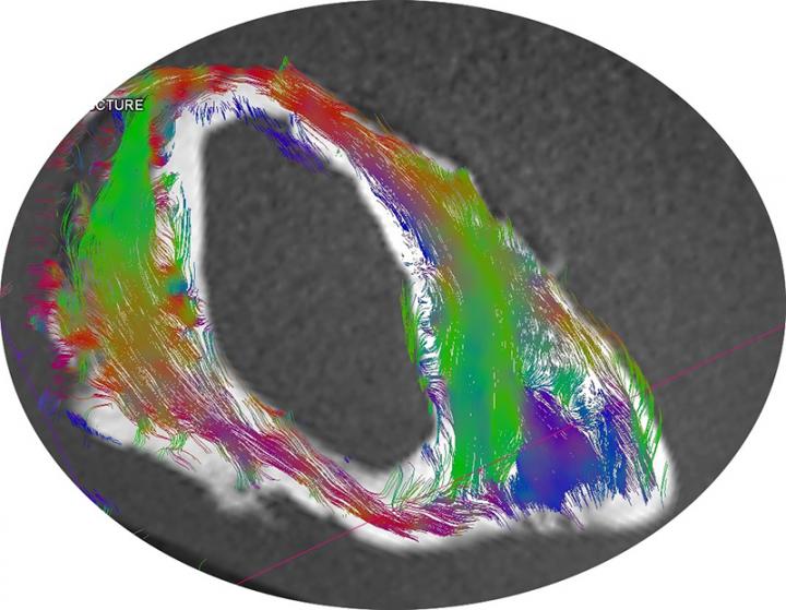
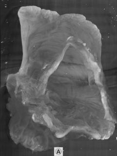
Edinburgh Imaging has been collaborating with the equine cardiology team at the RDSVS to evaluate the myofibre pattern in the equine left atrium.


The equine cardiology team at the The Royal (Dick) School of Veterinary Studies were interested in evaluating the myofibre pattern in the equine left atrium.
Knowledge of fibre patterns could provide important information for newly developing electroanatomic mapping techniques in horses.
Diffusion tensor MR & micro CT scanning (using iodine contrast) have been successfully applied to cardiac tissue in other species to determine myofibre orientation.
The advantage of these techniques over traditional histopathology, is that they are non-destructive, thus anatomical features & relationships can be preserved.
With funding from the Horseracing Betting Levy Board, the equine cardiology team collaborated with Edinburgh Imaging & Edinburgh pre-clinical imaging teams to scan 22 post mortem equine / horse atrial specimens.
The results were encouraging, with excellent visualisation of the 3D spatial organisation of fibre tracts using both techniques - the equine cardiology team is currently analysing the data.
Social media tags & titles
Edinburgh Imaging has been collaborating with the equine cardiology team at the RDSVS to evaluate the myofibre pattern in the equine left atrium.
@TheDickVet @DickVetEquine @levyboard

