What is a MR scan?
Magnetic resonance (MR) imaging was first demonstrated in the early 1970's & first used clinically in the 1980's. It is a relatively new technique which continues to be a fast developing science.

What is a MR scan & what is it used for?
Magnetic resonance (MR) imaging uses strong magnetic fields & radio waves to produce detailed images of the inside of the body.
A MR scan can be used to examine almost any part of the body, including the:
- Brain & spinal cord
- Bones & joints
- Breasts
- Heart & blood vessels
- Internal organs, such as the liver, womb or prostate gland
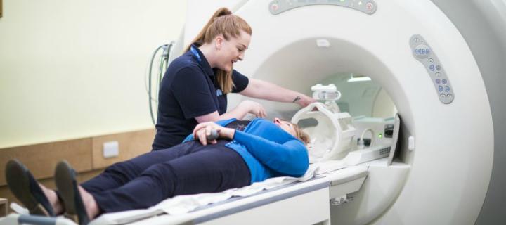
A MR scanner is a large tube that contains powerful magnets.
MR is unique in that it uses a combination of high field strength magnets & radio waves, as well as the magnetic properties of hydrogen in water, to create detailed pictures.
Most MR scanners have a 1.5 tesla superconducting magnet (1.5 T) - that is 30,000 times stronger than that of the earth's magnetic field!
You lie inside a tube during the scan.
The results of a MR scan can be used to help diagnose conditions, plan treatments & assess how effective previous treatment has been.
Relevant Edinburgh Imaging publications

