
27 Jul 17. Science Insights
Science Insights is an exciting work experience programme designed to give 5th year high school pupils a real insight into the work and life of research scientists.
The annual programme – launched in 2014 by the MRC Institute of Genetics & Molecular Medicine and The Roslin Institute – has proved a great success. The five research institutes in the College of Medicine and Veterinary Medicine at the University of Edinburgh have participated from 2016, enabling an increased number of places for pupils on the programme.
This year's programme will provide an opportunity for 40 high school pupils to spend a week of their summer holidays following a varied programme of activities on four different University of Edinburgh campuses, gaining a real insight into research and work in biological sciences, medicine and veterinary medicine.
Science Insights at the Bioquarter Campus
Edinburgh Imaging were delighted to welcome 12 of these high school pupils today, whilst they were visiting the Bioquarter Campus.
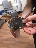
Each pupil received a tour of the Edinburgh Imaging RIE Facility, where our research radiographers explained that as the human body is mostly water, these water molecules (H2O) contain hydrogen nuclei (protons) which become aligned in a magnetic field. Sean and Lucy demonstrated how a magnet underneath a petri dish of ferrofluid forms spikes along the magnetic field lines.

Our radiographers then showed them our magnetic resonance imaging (MRI) scanner, and explained how it is often described as a doughnut-shaped magnet. This magnet is very strong and great care has to be taken to ensure that all moveable metal objects are away from the scanner before it’s switched on – Lucy demonstrated its strength by using a metal key!
Sean and Lucy discussed how the MRI is often used for neuroimaging research, such as small vessel disease, dementia and strokes. It can also be used to detect changes in your brain as you perform different tasks and experience different emotions. This application is called functional MRI (fMRI) and it works by detecting which parts of the brain are being supplied with higher levels of oxygenated blood compared to others. This can help neuropsychologists understand the function of different brain areas as well as helping the treatment of brain damaged patients.
Edinburgh Imaging - alot more than just MRI
The school pupils, then enjoyed a presentation by Jane on the Science of Imaging, describing
- the imaging modalities available in research institutes across Edinburgh, whether veterinary, clinical, biomedical or preclinical
- the purpose of xrays, ultrasound, computed tomography (CT), magnetic resonance imaging (MRI) scanner, positron emission tomography (PET) and microscopy
- the vast number of careers available within an imaging environment, such as becoming a medical physicist, image analyst, radiochemist or radiographer
Different careers in imaging
Shadia, a PhD student from Clinical Sciences, then explained her journey from an undergraduate in Computer Engineering to her current position as a part time PhD student, whilst working with Toshiba, developing software.
Hopefully the school pupils found it interesting to discover that there are lots of opportunities for people who haven't a traditonal medical undergraduate degree, to come and work at research institutes, such as the ones based in the Bioquarter campus.
We hope that they will have discovered that imaging is a vital part of health research, and through scientists using microscopy, MRI, CT scans and PET imaging, they are advancing health through excellence in imaging science.