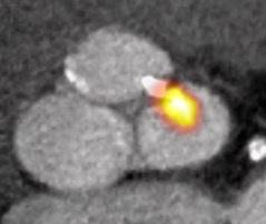
Aortic stenosis
Aortic stenosis is a problem whereby the exit from the heart’s left ventricle into the aorta, is narrowed & may affect the valve separating the two structures, or the tissues above or below the valve.

Overview
In patients with aortic stenosis, we demonstrated that the electrocardiographic strain pattern associated with left ventricular hypertrophy is indicative of myocardial fibrosis. We described a risk prediction model for patients with aortic stenosis demonstrating the important role of myocardial fibrosis imaging in determining future events.
This formed the basis of the Sir Jules Thorn Award for Biomedical Research 2015 to Dr Marc Dweck, which has funded a multi-centre clinical trial of early surgery in patients with advanced asymptomatic aortic stenosis using MR-based T1-mapping techniques, to characterise the fibrosis progression associated with this disease.
Lead aortic stenosis researcher
To discuss new research & collaborative imaging projects with Edinburgh Imaging, please contact:
Edinburgh Imaging
Enquiries: studies / collaborations / facilities
Contact details
- Email: edimg.studyinfo@ed.ac.uk
Research staff with an aortic stenosis focus
Current projects
Completed projects
Funding organisations & groups
Organisations are listed alphabetically:
Relevant links
Relevant Edinburgh Imaging publications
-
18 Feb 21. Featured Paper. Contrast-enhanced computed tomography assessment of aortic stenosis.
-
13 Feb 19. Featured Paper. Imaging & impact of myocardial fibrosis in aortic stenosis.
-
21 Aug 18. Featured Paper. Cardiac CT in prosthetic aortic valve complications.
- 04 Apr 18. Featured Paper. Computed tomography aortic valve calcium scoring in patients with aortic stenosis.
- Please view all our publications, here

