Heart
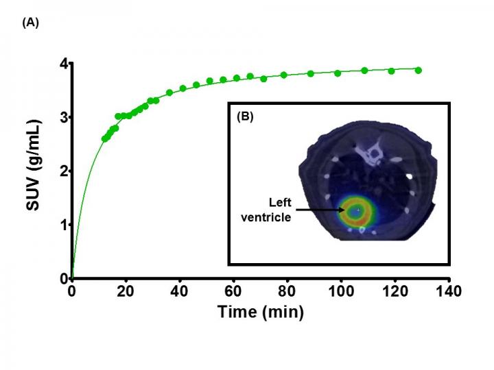
Example of cardiac PET/CT study in rats.
Evaluating myocardial viability using 18F-FDG and microPET/CT imaging.
In this study, 18F-FDG PET was used to evaluate myocardial viability in a rat model of myocardial infarction. Representative cardiac-gated PET images obtained following intravenous bolus injection of 18F-FDG in adult female Sprague-Dawley rats are shown below.
Dynamic microPET/CT images of rats’ myocardium were obtained over 2 hours post injection of 18F-FDG. Time-activity curves were obtained and kinetic modelling performed using image derived input functions for quantification of myocardial glucose metabolic rate (see figure below). (A) Radioactive concentration measured over time in a rat left ventricle following intravenous bolus injection of 18F-FDG. (B) microPET image showing radiotracer uptake in the left ventricle co-registered with microCT image of same animal.


