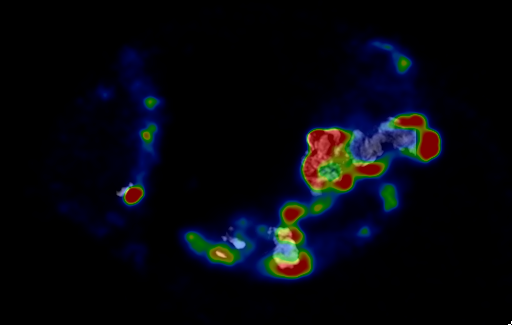
Human aortic valve
Using microPET/CT images to understand the mechanisms underlying calcification of human aortic valves.

Our preclinical PET/CT scanner is capable of acquiring images with a much higher sensitivity and resolution than a clinical PET scanner, allowing in-depth analysis of oartic calcification in human tissues. (A) MicroCT image co-registered to microPET image demonstrating mismatch between 18F-NaF uptake distribution and CT density in a severely calcified human aortic valve. (B) Volume rendering of same human aortic valve shown in (A).

