SEM imaging applications
The Scanning Electron Microscope (SEM) supports a wide variety of diverse applications including secondary electron imaging, backscatter electron imaging, cathodoluminescence imaging and electron backscatter diffraction.
For further details about imaging and sample requirements please contact our facility staff.
Secondary Electron (SE) Imaging
Secondary electrons produced when an electron beam interacts with the surface of the sample, enable fine-scale (nm) surface features and topographic detail to be imaged. The SEM images have far greater spatial resolution and depth of focus than optical microscopes.
Backscatter Electron (BSE) Imaging
Backscatter electrons allow compositional variation to be imaged as different grey-scales. Materials with a mean high atomic number composition appear brighter than those of low atomic number, enabling the imaging of compositional variation and growth zoning.
Cathodoluminescence (CL) imaging
Certain materials will emit light when bombarded by an electron beam. This luminescence can be imaged using a CL detector. Variations in the intensity of the light emitted can be influenced by impurities and crystal defects. CL is particularly useful for imaging features such as growth zoning or crystal overgrowths that are not easily imaged by other methods.
Electron Backscatter Diffraction (EBSD)
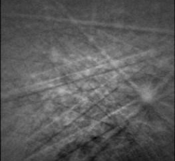
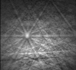
EBSD can be used to obtain crystallographic information from the diffraction patterns generated when the electron beam interacts with a crystalline sample. The technique can provide information for absolute crystallographic orientation, microtextures and preferred crystal orientation, deformation and strain, polymorph identification, damage to crystal structure, grain size and grain boundaries.
The technique can be used to analyse:
- Absolute crystallographic orientation
- Microtextures and preferred crystal orientation
- Deformation and strain
- Polymorph identification
- Damage to crystal structure
- Grain size and grain boundaries
Damage to crystal structure
We have provided 'An EBSD study of the damage to zircon crystals' by Dr Nicola Cayzer and Dr Richard Hinton from the School of GeoSciences.
It is available for PDF download:
Identification of polymorphs
The EBSD technique can be used to identify different polymorphs from their diffraction patterns.

Silica inclusions within diamond can readily be identified as monoclinic coesite or trigonal quartz by in situ, non-destructive point analyses.

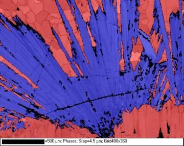
The distribution of aragonite and calcite within speleothems, bivalves and corals can be mapped with a resolution of ~1 µm. This image shows aragonite needles (blue) in a speleothem surrounded by calcite (red).


