26 Oct 22. Breast cancer detection
Is dynamic enhancement of breast lesions on contrast enhanced spectral mammography (CESM) possible at sub-lesion level: exploring registration techniques to allow sub-lesion analysis?

We presented pilot results from a project investigating the detection of breast cancer at the recent Cambridge Conference on Breast Cancer Imaging 2022.
Background
The collaborative work between Dr Calum Gray (Edinburgh Imaging), and Dr Sarah Savaridas (School of Medicine, University of Dundee) initially came about when Dr Savaridas, who is a Consultant Radiologist at NHS Tayside, posted on the SINAPSE Technical MR forum for advice on analysing breast mammography images. Dr Gray from the Image Analysis Team responded with some ideas, which led to further discussions and setting up of this project.
Contrast enhanced spectral mammography (CESM) is a functional imaging technique demonstrating lesion vascularity through contrast uptake. CESM has similar diagnostic accuracy to breast MR, whilst also being quicker and more acceptable to patients. However, in addition to morphological data, it is possible to derive time intensity curve (TICs) from MR images which provide greater functional information. Analysis is performed at sub-lesion level to identify the most aggressive features in heterogeneric breast cancers.
This is the first study to explore the possibility of aligning early and delayed CESM MLO views to allow similar sub-lesion dynamic enhancement analysis.
What we did
CESM images were acquired as part of prospective breast imaging studies, CONTEST and CONDOR at the University of Dundee. The initial MLO of the index breast was acquired three minutes after contrast injection. A delayed MLO view was acquired after a delay of six minutes. Fifteen subjects were selected at random from the cohort.
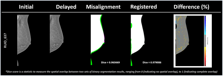
The Image Analysis team used a combination of rigid and non-rigid registration techniques to spatially align the two MLO views, create subtraction images, and then overlay colour maps for visual interpretation.
Outcomes
The study showed that registration was achieved in all 15 cases, with good agreement shown by both visual assessment and Dice similarity coefficient.
The significance of the project is that it is the first study to demonstrate that it is possible to align early and delayed MLO views to allow dynamic enhancement features to be demonstrated via colour mapping. This will allow sub-lesion analysis of breast cancer on CESM studies, which will ultimately improve the diagnostic and treatment pathway for patients.
The team are now seeking further funding to enable the process to be applied in a larger cohort of mammography scans.
Contact Dr Sarah Savaridas for more information. s.savaridas@dundee.ac.uk
Relevant links
Social media tags and titles
Is dynamic enhancement of breast lesions on contrast enhanced spectral mammography (CESM) possible at sub-lesion level: exploring registration techniques to allow sub-lesion analysis?

