
Personal profile
- 2017-present: Postdoctoral Research Fellow, University of Edinburgh
- 2014-2016: Marie Curie Postdoctoral Fellow, University of Edinburgh
- 2014: Embryology Course student, Woods Hole, Massachusetts
- 2008 - 2013: PhD in Developmental Neurobiology, BRAINSHARK group, University of Santiago de Compostela, Spain
- 2007 – 2008: Postgraduate Program in Neuroscience, University of Santiago de Compostela, Spain
- 2001-2007: Bachelor’s Degree in Biology, University of Santiago de Compostela, Spain
- 2005-2007: Specialization in Molecular Biology, University of Santiago de Compostela, Spain
- 2004-2005: Specialization in Biotechnology and Genetics, University of Lisbon, Portugal
Other roles:
Member of Biomedical Sciences Opportunities Committee (BSOC)
Member of George Square Postdoctoral (G2PD) Committee
Early Career Reviewer for Elife Journal
Preprint pool of selectors for Prelights (The Company of Biologists)
https://prelights.biologists.com/profiles/idoia-qu/
Students supervision: 1 PhD student, 4 master students, 1 honours student

Research
I am a developmental neurobiologist (with background in evo-devo) interested in understanding how our brains form during embryonic development.
I have experience and interest in brain embryonic development of different species, from sharks to humans and I am fascinated about their similarities and uniqueness. My brain areas of expertise are the telencephalon and the diencephalon, the embryonic structures that give rise to adult structures such as the cortex, thalamus or basal ganglia.
I believe that only through the study of evolution we will be able to achieve a full understanding of any biological process including, of course, the incredibly complex task of creating an adult brain from a limited starting number of progenitor cells.

In my current project I am using human induced pluripotent stem cells (hiPSCs) to investigate potential developmental causes of autism spectrum disorder.
Techniques:
Transcriptomics: Single cell and bulk RNAseq
Bioinformatics
Human induced pluripotent stem cell culture
In vitro differentiation (2D/3D) of inhibitory and excitatory neurons
Embryonic brain slice culture and heterotypic explant assays
Axon guidance assays
Tract-tracing techniques (DiI, Neurobiotin)
Cell cycle analysis (BrdU, EdU)
Luciferase assays
Immunohistochemistry, in situ hybridization, qPCR, general histology
Experimental models:
Shark (Scyliorhinus canicula) embryos, mouse, human pluripotent stem cells.
Relevant publications
*Co-corresponding authors
Quintana-Urzainqui I*, Hernández-Malmierca P, Clegg JM, Li Z, Kozić Z, Price DJ. (2020) The role of the diencephalon in the guidance of thalamocortical axons in mice. Development. DOI:10.1242/dev.184523
Quintana-Urzainqui I*, Kozić Z,Mitra S, Tian T, Manuel M, Mason JO, Price DJ. (2018). Tissue-Specific Actions of Pax6 on Proliferation and Differentiation Balance in Developing Forebrain Are Foxg1 Dependent. iScience. 10:171-191. DOI: 10.1016/j.isci.2018.11.031
Docampo-Seara A, Lagadec R, Mazan S, RodrÃguez MA, Quintana-Urzainqui I*, Candal E*. (2018) Study of pallial neurogenesis in shark embryos and the evolutionary origin of the subventricular zone. Brain Struct Funct. 223:3593-3612. DOI: 10.1007/s00429-018-1705-2
Santos-Durán GN, Ferreiro-Galve S, Menuet A, Quintana-Urzainqui I, Mazan S, Rodríguez-Moldes I, Candal E. (2016). The Shark Alar Hypothalamus: Molecular Characterization of Prosomeric Subdivisions and Evolutionary Trends. Front Neuroanat. 10:113.
Caballero I, Manuel MN, Molinek M, Quintana-Urzainqui I, Mi D, Shimogori T, Price DJ. (2014). Cell-autonomous repression of Shh by transcription factor Pax6 regulates diencephalic patterning by controlling the central diencephalic organizar. Cell Rep. 11:1405-1418.
Quintana-Urzainqui I., Rodríguez-Moldes I., Mazan S., Candal E. (2014). Tangential migratory pathways in the developing telencephalon of sharks: evolutionary implications. Brain Struct Funct 2014. DOI: 10.1007/s00429-014-0834-5
Quintana-Urzainqui I, Anadón R, Candal E, Rodríguez-Moldes I. (2014). Development of the Terminal Nerve System in the shark Scyliorhinus canicula. Brain Behav Evol. 84:277-287.
Quintana-Urzainqui I., Rodríguez-Moldes I., Candal E. (2014). Developmental, tract-tracing and immunohistochemical study of the peripheral olfactory system in a basal vertebrate: insights on Pax6 neurons migrating along the olfactory nerve. Brain Struct Funct. 219:85–104
Quintana-Urzainqui I., Sueiro C., Carrera I., Ferreiro-Galve S., Santos G., Pose-Méndez S., Mazan S., Candal E., Rodríguez-Moldes I. (2012). Contributions of Developmental Studies in the Dogfish Scyliorhinus canicula to the Brain Anatomy of Elasmobranchs: Insights on the Basal Ganglia. Brain Behav Evol. 80:127-141.
Rodríguez-Moldes I., Carrera I., Pose-Méndez S., Quintana-Urzainqui I., Candal E., Anadón R., Mazan S., Ferreiro-Galve S. (2011). Regionalization of the shark hindbrain: a survey of an ancestral organization. Front Neuroanat. 5: 16.
Carrera I., Anadón R., Quintana-Urzainqui I., Pose-Méndez S., Rodríguez-Moldes I. (2010). Development of descending supraspinal pathways in a shark and neurochemical characterization of projection neurons. Int J Dev Neurosci. 28: 668-669.
Book chapter:
Rodríguez-Moldes, I., Santos-Durán, G.N., Pose-Méndez, S., Quintana-Urzainqui, I., Candal, E., (2017). The Brains of Cartilaginous Fishes. In: Kaas, J (ed.), Evolution of Nervous Systems 2e. vol. 1, pp. 77–97. Oxford: Elsevier.
Journal cover:
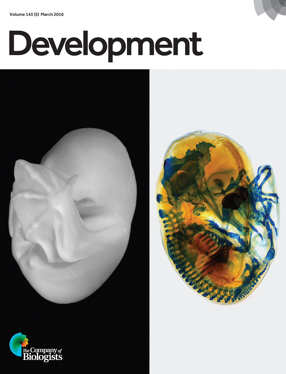
Development cover (Volume 143 (5) March 2016) http://thenode.biologists.com/wp-content/uploads/2016/12/5.jpg