
Images
Chicken embryo and related images
Our Creative Commons images are free to download for non-commercial use. If you use any of our photos, please add the credit line "Photo courtesy of The Roslin Institute Chicken Embryology group (RICE), The Roslin Institute, The University of Edinburgh". If you have an Acknowledgements section, please add “Photo courtesy of The Roslin Institute Chicken Embryology group (RICE), The Roslin Institute, The University of Edinburgh, UK.
Click on the title link next to each image to access the full-size version for download.
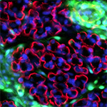 |
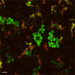
Chimeric muscle |
Section through breast muscle of chimeric embryo derived from GFP-expressing donor and wild-type host. Muscle collected from E12.5 embryo and section immunostained for laminin (red) and dapi (blue). Image is x600. |
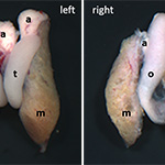 |
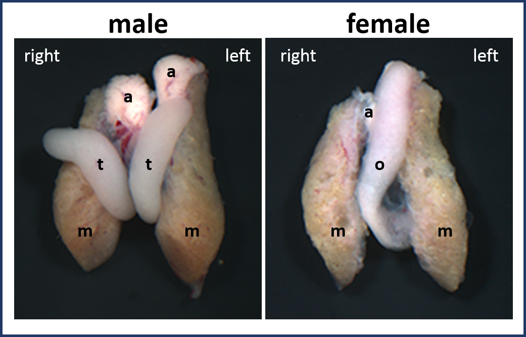
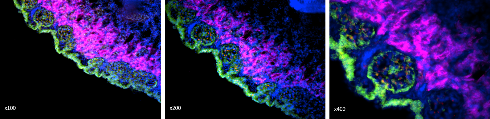
Embryonic chick gonads |
Light microscopy images of gonads from E19 male and female chick embryos. In the male embryo the two testes have a ‘tubular’ morphology and are of similar size. In the female, the left ovary is a larger structure with a ‘ribbon-like’ morphology (the right ovary is regressing). Image caption: [t=testis; o=ovary; m=mesonephros; a=adrenal]. |
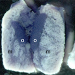 |
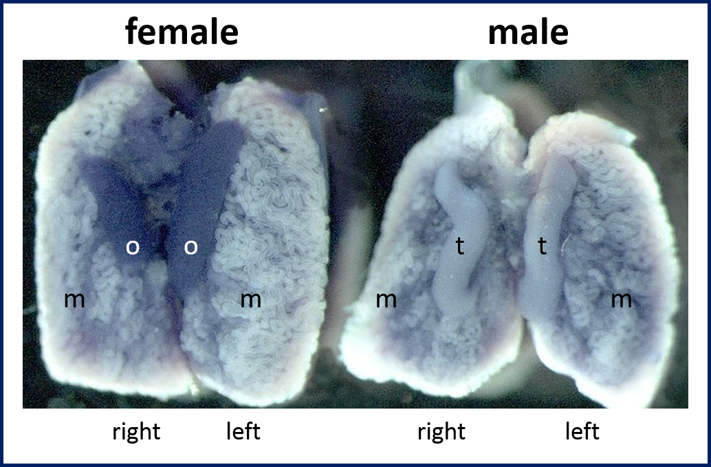
Faf expression in embryonic chick gonads |
In situ hybridisation of Faf transcript in E9.5 female and male chick gonads. Sites of Faf expression is shown by blue stain. Image caption: [t=testis; o=ovary; m=mesonephros] |
 |
Ovarian germ cell cysts | Somatic cell capsules containing germ cells/ presumptive oocytes in the cortex of an embryonic chick ovary just before hatch. |
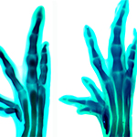 |
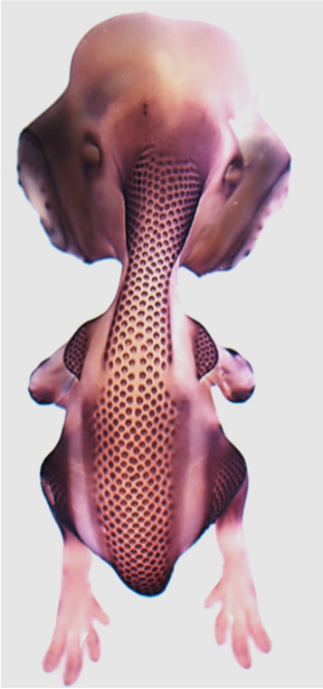
Chicken feet normally develop 4 toes, however several breeds of chickens develop an extra toe on the anterior side of the foot (the ‘big toe’ side). This is due to a mutation in a long range limb enhancer of Sonic hedgehog, which causes excessive SHH protein to be made. Similar mutations cause polydactyly in humans, cats, dogs and mice. |
|
 |
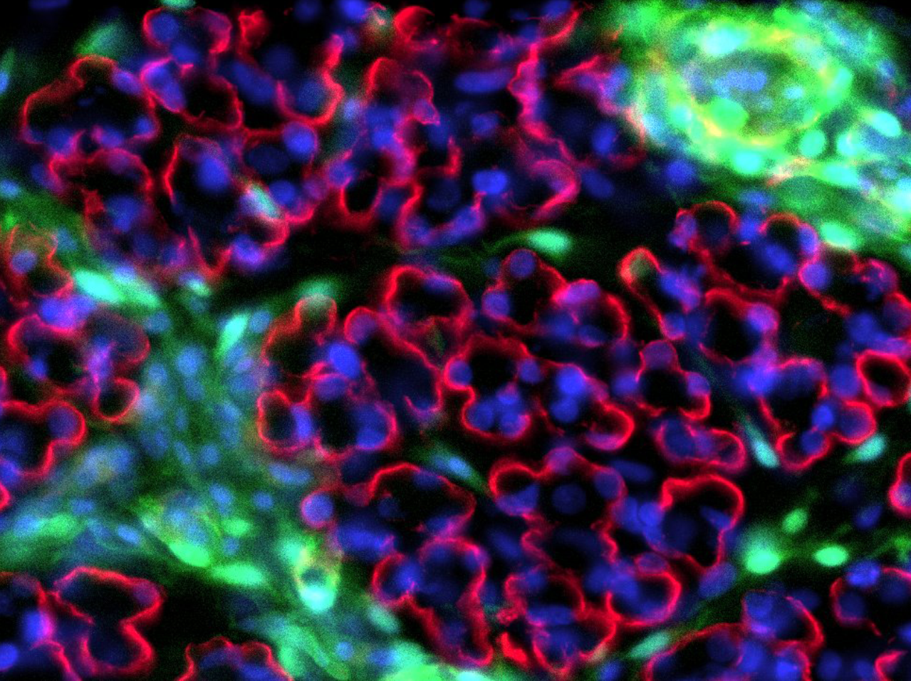
CSF1R-eGFP transgene expression highlights lymphoid structures |
CSF1R-eGFP (green) transgene expression in the follicle-associated epithelium of the bursa of Fabricius. F-actin (red) and jacalin lectin (blue) staining also shown. |
 |
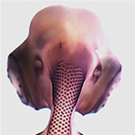
Images are from embryonic stages (E)5-7. Note optic fissure margin (OFM) at ventral aspect of the eye. The Rainger lab has been set up to identify the molecular processes which guide the closing of the OFM, to help identify new genetic drivers of this event. Such information will help understand the causes of ocular birth defects, such as Coloboma, as well as contributing to insights into other fusion processes (e.g. palate, neural tube, heart, diaphragm) or wound healing. Scale bar = 1 mm. L, lens; NR, neural retina. |
|
 |
Dorsal view of chicken embryo showing regions of feather follicle formation. Purple signal indicates expression of CTNNB1 as revealed by in situ hybridisation. |
|
 |
Fluorescent image of cultured skin from dorsal side of GFP-chicken showing feather follicles condensing in waves from midline outwards. |
|
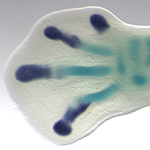 |
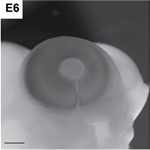
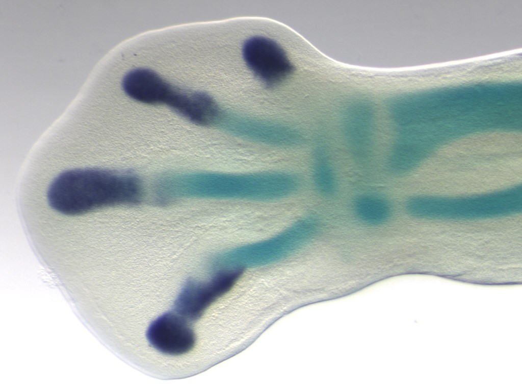
RNA in situ hybridisation showing expression of SOX9 in an embryonic chicken foot (dark blue), and which marks cells destined to become cartilage cells. Alcian green shows cells which have already become cartilage (and turned off SOX9). |
|
 |
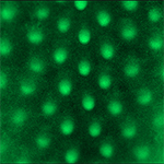
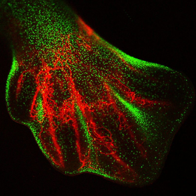
Macrophages (green) are concentrated in areas of cell death, blood vessels (red) are labeled with SNA lectin. |
|
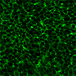 |
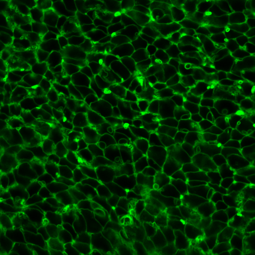
Fluorescent image of next-to-outer layer of dorsal skin from membrane-GFP chick embryo. |
|
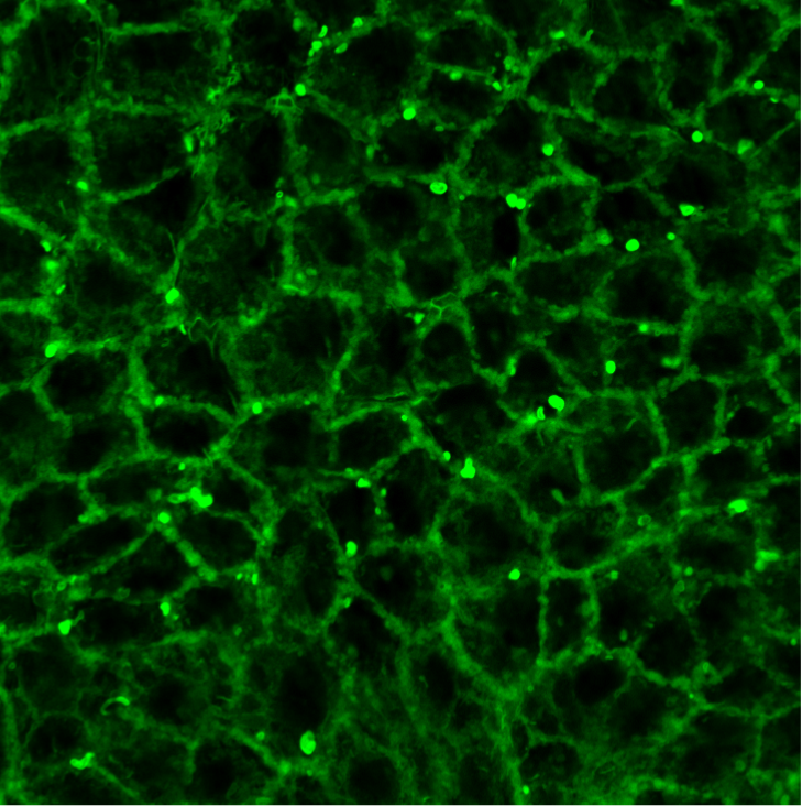
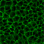 |
Fluorescent image of the most outer layer of dorsal skin from membrane-GFP chick embryo. |
|
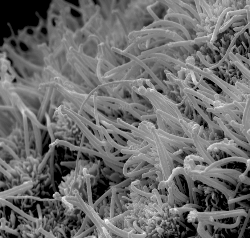
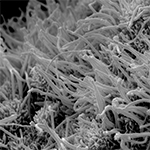 |
Motile cilia are important for movement of liquids in the body. In many tissues they beat synchronously, such as in the lung and trachea. Motile cilia first develop on the ependymal cells of the brain ventricles at E8 during chicken embryonic development where they move cerebrospinal fluid. Each cell develops multiple cilia. |
|
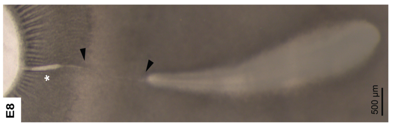
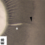 |
Superficial closure of the OFM is complete in the neural retina region (between arrows) of the chick eye by E8. Note the iridial region maintains an opening (asterisk). We are investigating this closure at a tissue and histological level, using transgenic tools such as the memGFP chick line. |
|
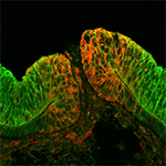 |
Validation by immunoflourescence of a OFM candidate gene (red) identified by RNAseq. Protein is localised to cells within the opposing sides of the fissure margin prior to closure in an E6 chick eye. N-cadherin is used as a counterstain (green) and shows neural-retina specific localisation. |
|
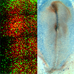 |
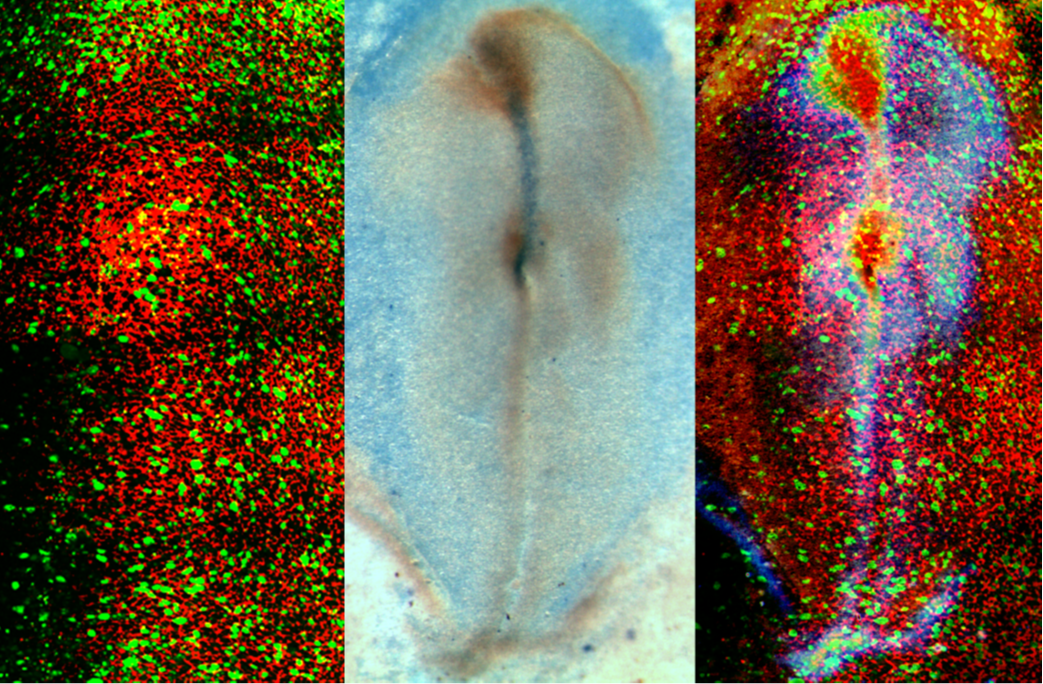
In situ hybridisation and fluorescent imaging of Stage 5HH- chicken embryo primitive streak stage gastrulation. Sonic Hedgehog (SHH-blue stain) is expressed asymmetrically in Hensen’s node, the early organiser of Left/Right asymmetry. Posterior (bottom of the picture) the primitive streak can be seen, as a central midline stripe, which does not stain. Here, cells of the epiblast are ingressing through the furrow of the primitive stream and becoming either mesoderm or endoderm. Anterior (top of the picture) SHH is expressed in the notochord (a single blue line), a mesodermally derived structure which will become the organiser of the neural plate. The embryo is said to ‘break symmetry’ at the node- and after gastrulation cells know if they are on the right or the left hand side of the embryo. VANGL2 is a protein in the Planar Cell Polarity pathway (PCP).After gastrulation cells on either side of the primitive streak, which have ‘broken symmetry’ localise the VANGL2 within the cell to the side of the cell which faces the primitive streak. |
|
 |
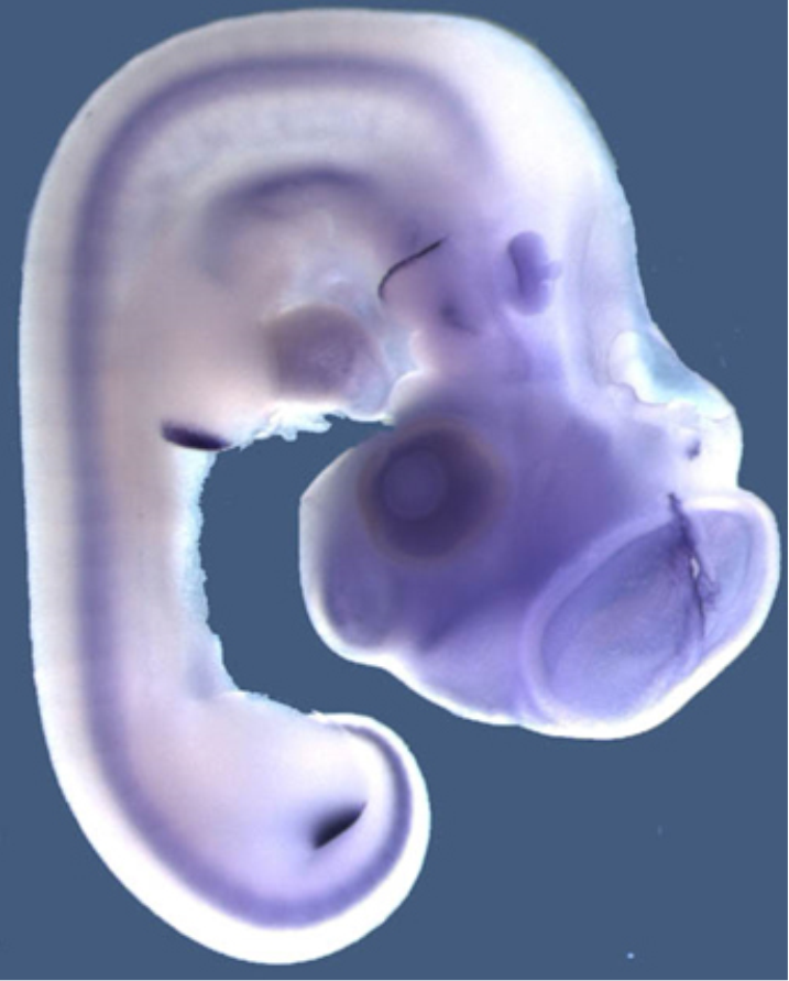
SHH is expressed in many ‘organiser’ tissues during chicken development (blue). These tissues use SHH as a signal to communicate to the other tissues around then and instruct them to take on specific identities. For example SHH is expressed in the posterior limb bud which communicates to the limb bud tissue the number and identity of digits which form. |
|
 |
Multiple transgenes highlight different immune cell populations |
Cells in the embryonic yolk-sac expressing the RUNX1-eGFP (green) or CSF1R-mApple (red) transgenes. |