Instant scans give insight into disease
A unique imaging facility will help researchers and doctors improve diagnosis and treatments for a range of illnesses.
The £20 million Clinical Research Imaging Centre (CRIC) is the first fully integrated imaging facility of its kind in the UK.
The centre enables research into illnesses including multiple sclerosis, schizophrenia, cancer and heart disease.
It is a collaboration between the University and NHS Lothian and was officially opened by HRH, The Duke of Edinburgh.
Video report
Watch a report on the Clinical Research Imaging Centre.
Use of imaging
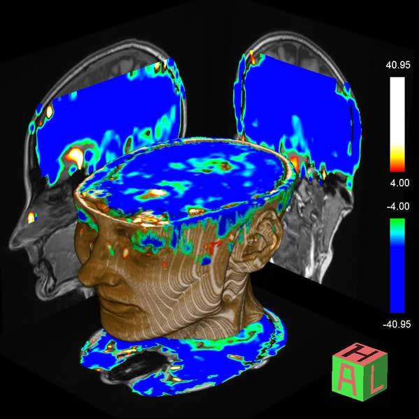
The different scanners at the centre can be used study the causes and spread of diseases on different ways and assess their impact on the body.
The scanners allow investigations to take place without invasive procedures.
Their use reduces the need for biopsies or angiograms, where catheters are used to identify vessel and organ damage.
Experts can use the imaging equipment to scan organs in under a second and to see in great detail how they function.
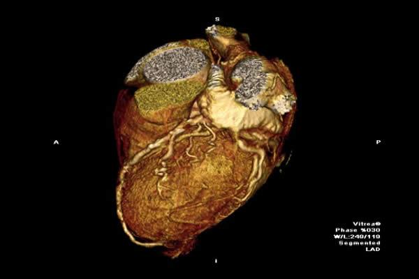
They are also also track the flow of blood through vessels, for instance in the heart, determine the spread of disease and assess the effectiveness of new drugs treatments.
As opposed to simply looking at the structures of the body – such as the heart and the brain – we can look at how organs are functioning in real time. This will not only help us better understand disease but it will help us to improve both diagnosis and treatments.
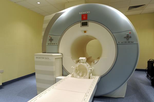
Thanks to our supporters we've been able to fund the MRI scanner in this fantastic new centre in Edinburgh. The machine will help researchers to uncover vital new insights about the heart and offer cutting-edge treatment to patients at the Royal Infirmary.
In pictures
Scanners
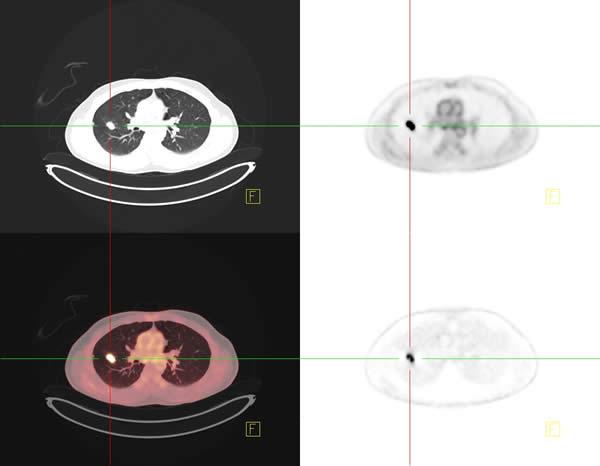
CRIC houses a high strength magnetic resonance imaging (MRI) scanner, the world's most advanced computerised tomography (CT) scanner and a CT-positron tomography scanner.
A cyclotron and radiochemistry laboratories on-site can also produce tracer chemicals that can be used to investigate the spread of cancer.
Tracer chemicals can also be produced to investigate inflammation in tissues.
Such inflammation is a key factor in heart and lung diseases as well as changes in the brain in diseases such as multiple sclerosis and Parkinson’s disease.


Funding
Supporters of the Clinical Research Imaging Centre include
- Medical Research Council
- Wellcome Trust
- British Heart Foundation (BHF)
- European Regional Development Fund
- Scottish Funding Council
- Private organisations and individuals
This world-leading new centre brings together the very latest imaging technologies in a single facility. With the University of Edinburgh’s world-leading clinical research, this will allow a major improvement in our ability rapidly to investigate and understand the most serious and distressing diseases in our patients

