Eyes / retinal
Are eyes a window to the brain's health?

The retina is an extension of the brain, sharing embryonic origins as well as features such as nerve tissue, small blood vessels & a blood-tissue barrier; it therefore has the potential to reveal important aspects of brain health.
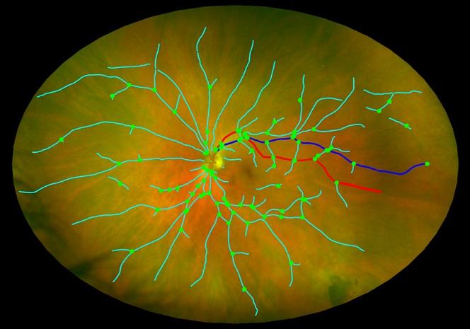
The retina is the light-sensing inner surface of the human eye & is comprised of layers of tissue consisting of neurons & supporting cells, interconnected by synapses. The retinal ganglion cell (RGC) layer, the innermost cellular layer, projects its axons across the inner retina, called the retinal nerve fibre layer (RNFL), through the optic nerve to the visual processing & cognitive centres of the brain. The retina is unique in the human body in allowing easy observation of blood vessels with simple, non-invasive optical instruments such as the fundus camera. Evaluating the health of blood vessels is crucial to studying various diseases which affect both the body & brain.
Our retinal imaging team is partnered with NHS Lothian & researchers at The University of Edinburgh to investigate associations between features measured on retinal imaging & diseases such as diabetes, hypertension, liver disease / cirrhosis, kidney disease, stroke, multiple sclerosis, small vessel disease, cardiovascular disease & dementia. The impetus for these investigations is primarily the need for new biomarkers capable of improving the identification of susceptible individuals, acting as early indicators of illness, aiding disease characterisation & prognosis, & assessing the efficacy of new treatments.
Edinburgh Imaging staff with a focus in retinal imaging
- Dr Neeraj Dhaun
- Prof Baljean Dhillon
- Dr Fergus Doubal
- Prof Jonathan Fallowfield
- Dr Tom MacGillivray
- Prof Craig Ritchie
- Prof Joanna Wardlaw
- Dr Stewart Wiseman
Current projects
Completed projects
Funding organisations & research groups
Listed alphabetically:
- Alzheimer’s Drug Development Foundation (ADDF)
- Alzheimer’s Research UK Scotland Network
- EPSRC
- Innovate UK
- Medical Research Council (MRC)
Relevant Edinburgh Imaging publications

