11 Dec 20. Featured Paper
Automated segmentation of optical coherence tomography angiography images: benchmark data & clinically relevant metrics.

Link to paper on Translational vision science & technology.
Authors
Ylenia Giarratano; Eleonora Bianchi; Calum Gray; Andrew Morris; Tom MacGillivray; Baljean Dhillon; Miguel O. Bernabeu
Abstract
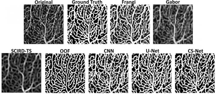
Purpose: To generate the first open dataset of retinal parafoveal optical coherence tomography angiography (OCTA) images with associated ground truth manual segmentations, & to establish a standard for OCTA image segmentation by surveying a broad range of state-of-the-art vessel enhancement & binarization procedures.
Methods: Handcrafted filters & neural network architectures were used to perform vessel enhancement.
Thresholding methods & machine learning approaches were applied to obtain the final binarization.
Evaluation was performed by using pixelwise metrics & newly proposed topological metrics.
Finally, we compare the error in the computation of clinically relevant vascular network metrics (e.g., foveal avascular zone area & vessel density) across segmentation methods.
Results: Our results show that, for the set of images considered, deep learning architectures (U-Net & CS-Net) achieve the best performance (Dice = 0.89).
For applications where manually segmented data are not available to retrain these approaches, our findings suggest that optimally oriented flux (OOF) is the best handcrafted filter (Dice = 0.86).
Moreover, our results show up to 25% differences in vessel density accuracy depending on the segmentation method used.
Conclusions: In this study, we derive & validate the first open dataset of retinal parafoveal OCTA images with associated ground truth manual segmentations.
Our findings should be taken into account when comparing the results of clinical studies & performing meta-analyses.
Finally, we release our data & source code to support standardization efforts in OCTA image segmentation.
Keywords
- Optical coherence tomography angiography (OCTA)
Social media tags & titles
Featured paper: Automated segmentation of optical coherence tomography angiography images: benchmark data & clinically relevant metrics. @ARVOtvst @giaylenia @mobernabeu @TomJMacg #OCTA

