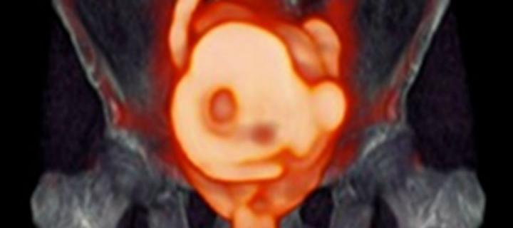
What is a PET scan?
Positron emission tomography (PET) scans are used to produce detailed three-dimensional images of how the cells are working inside the body.

What is a PET scan & what are they used for?
The detailed 3-dimensional images produced by a PET scan, can clearly show the part of the body being investigated, including any unusual areas, & can highlight how well specific functions of the body are working.
PET scans are often combined with CT scans to produce an even more detailed image. This is known as a PET-CT scan. They may also be combined with an MRI scan. This is known as a PET-MRI scan.
PET scans can be used to help diagnose a range of different cancers & can show how far a cancer has spread or how well it is responding to treatment. They can also be used to help diagnose a number of conditions that affect the normal workings of the brain (neurological conditions), such as dementia.
In the news

