Examples of Cardiac Ultrasound studies
Mouse
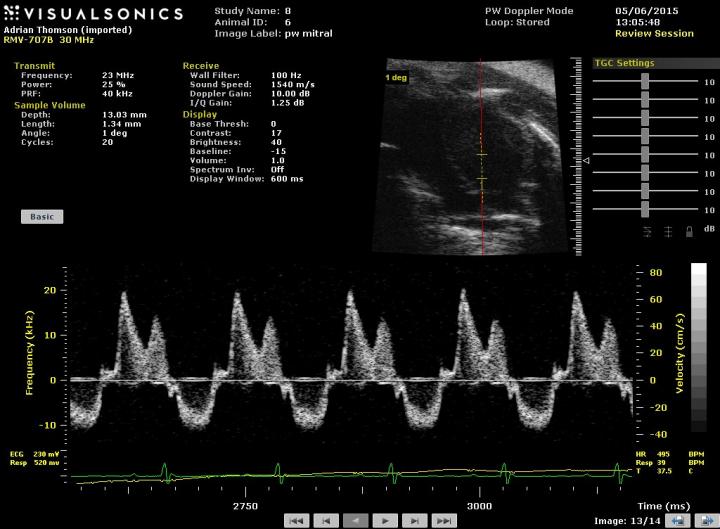
Spectral Doppler blood flow from normal adult mouse heart showing in-flow and out-flow of blood from 4-chamber view of normal mouse heart – image
Normal parasternal long-axis of adult mouse heart with left ventricle outlined and functional measurements displayed – video
E17.5 embryonic mouse heart showing left and right ventricles and valves – video
P1 neonate showing left ventricle of heart
Zebrafish
End systolic and end diastolic length and area measurements of an adult zebrafish heart
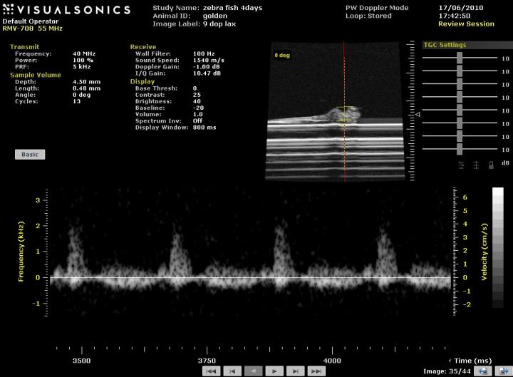
Spectral Doppler trace from a 4day post-fertilisation zebrafish embryo embedded in agar
References
White CI, Jansen MA, McGregor K, Thomson A, Richardson RV, Mylonas KJ, Moran CM, Seckl JR, Walker BR, Chapman KE, Gray GA. Cardiomyocyte and vascular smooth muscle independent 11β-hydroxysteroid dehydrogenase 1 amplifies infarct expansion, hypertrophy and the development of heart failure following myocardial infarction in male mice. Endocrinology. 2016 Jan; 157(1): 346-57.
Moran CM, Thomson AJ, Rog-Zielinska E, Gray GA. High-resolution echocardiography in the assessment of cardiac physiology and disease in preclinical models. Exp Physiol. 2013 Mar;98(3):629-44.
Gray GA, White CI, Thomson A, Kozak A, Moran C, Jansen MA. Imaging the healing murine myocardial infarct in vivo: ultrasound, magnetic resonance imaging and fluorescence molecular tomography. Exp Physiol. 2013 Mar;98(3):606-13.


