Examples of Studies using our Optical Imaging Equipment
Studies using our Optical Imaging Equipment
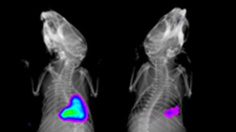
Detecting enzyme activity in vivo
A range of reagents are now available that fluoresce only in the presence of specific enzymes such as neutrophil elastases or cathepsins. Quantitative real time in vivo data such as this is not affected by processing artefacts present in ex vivo techniques and is ideal for time course studies.

Cathepsin sensitive probe in mice with lung injury in untreated (A) and treated (B). Images courtesy of Dr Kev Dhaliwal, CIR,QMRI.
Tracking Pathogens
Off the shelf bacterial strains can now be purchased which have the capacity to express light. These allow identification of the location and relative health of a bacterial population in vivo. These have been used widely in the literature for the testing of antibiotics and novel antimicrobial compounds.

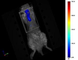
3D reconstruction. Labelled macromolecule accumulation in inflamed airway. Images courtesy of Dr Kev Dhaliwal, CIR,QMRI.
Tracking cells or Macromolecules
Cells or molecules can be fluorescently labelled prior injection into a host. An invaluable technique in the analysis of cellular migration and confirmation that your agent of interest reaches your region of interest. Luminescent and fluorescence expressing cells and cell lines are used widely in the study of cancer growth and invaluable for the accurate non invasive measurement of tumour size in drug trial programmes.
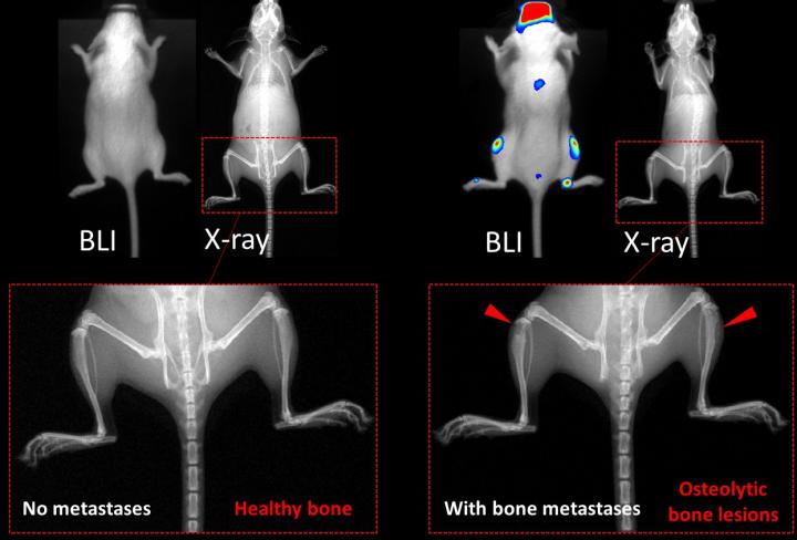
Experimental breast cancer bone metastasis model
Breast cancer bone metastases are generated by injecting cancer cells into the left ventricle of FVB mice. Breast cancer cell infiltration into bone marrow interferes with normal bone remodelling by increasing bone resorption, which causes fracturing.

In vivo imaging: bone metastatic burden on Day 21 post-intracardiac injection
Image courtesy of Meghan Maslen, 2nd Year PhD, Pollard and Qian Lab, CRH
For more examples or to discuss you project, email Paul Fitch or Adrian Thomson

