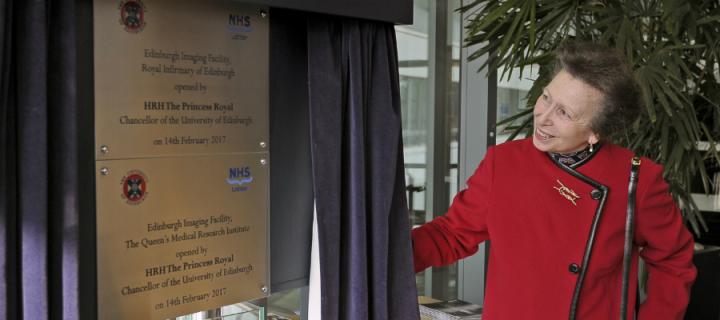
Chancellor opens new imaging suites
Feb 2017: Two advanced medical scanners have been installed as part of a £14 million makeover of research imaging facilities at the University of Edinburgh.
Edinburgh Imaging now houses six scanners dedicated to research, making it one of the largest networks in Europe with ties to clinical care.
New scanners
A purpose-built unit to house one of the new machines – a high-powered MRI brain scanner – has been opened by the University’s Chancellor, Her Royal Highness The Princess Royal.
The new suite is located within the Royal Infirmary of Edinburgh at Little France, close to the University’s existing facilities, and was built in collaboration with NHS Lothian.

Brain research
Research carried out with the new scanner will examine the brain at all stages of life – from birth to old age.
Studies involving babies and young children aim to shed new light on the factors that affect healthy brain growth in early life.
These new scanners make it possible to see the smallest details in the brain, even while babies are sleeping, meaning that we can learn so much more about how the brain changes throughout life and see if new treatments are helping, for example to slow down brain damage in dementia.
Stroke studies
In a separate study, researchers are leading an international team to investigate disorders that affect the blood vessels in the brain, leading to dementia and stroke in later life.
Funding for the redevelopment has been provided by Wellcome, Edinburgh and the Lothians Health Foundation, Dunhill Medical Trust, Theirworld and the Muir Maxwell Epilepsy Research Trust.
First in Scotland
Another advanced scanner – the first of its kind in Scotland – was installed within Edinburgh Imaging’s existing facilities at Little France.
The device combines MRI with another type of imaging technology – positron emission tomography (PET) – in a single machine.
Advanced technology
Combining the two types of scan into one machine allows researchers to view structures of the brain and other organs in action inside a person in real time.
The PET-MRI scanner was funded by the Medical Research Council as part of the Dementias Platform UK Imaging Network and will be fully operational later this year.
The upgrade to our existing facilities offers exciting new opportunities to link structure and function of organ systems, and link our capabilities together with advanced image analysis tools to serve our many research centres throughout the University as well as our NHS patients.
Edinburgh Imaging
More than £35m has been invested in Edinburgh Imaging facilities to date.
In addition to six advanced scanners, there are also on-site facilities that produce tracer chemicals called isotopes that are used in medical scans.
Symposium
Edinburgh Imaging is hosting a Science Symposium on 30 June, when international experts will meet to discuss the latest advances in biomedical imaging and analysis.
Related links
Neuroimaging Sciences at the Centre for Clinical Brain Sciences


